Pop! goes the beetle

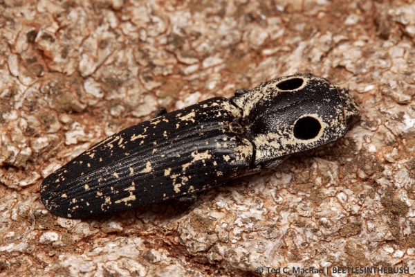
Alaus oculatus (eyed elater) | Beaver Dunes State Park, Oklahoma

Alaus oculatus (eyed elater) | Beaver Dunes State Park, Oklahoma

Last June while collecting beetles from cottonwood trees at Beaver Dunes State Park, Oklahoma, I came across one of my favorite beetles—Alaus oculatus, or eyed elater (family Elateridae, or click beetles). Large by click beetle standards, the most striking feature of eyed elaters is, of course, their false eye spots, which are not eyes at all but patches of pubescence—black surrounded by a narrow ring of white—intended to look like eyes and located prominently on the prothorax rather than the head. A handful of related species are also found in various parts of the U.S., all of which exhibit variations on this same eye spot theme. Undoubtedly these spots serve to frighten would-be predators, much like the false eye spots on the thorax of many lepidopteran caterpillars. The true eyes, of course, are much smaller and are located on the head in front of the “false eyes.” In contrast to the prominently visible eye spots, pubescence on the rest of the body seems to function in cryptic coloration. The mottled patterning blends in with the bark of trees where these beetles usually hang out for effective concealment.
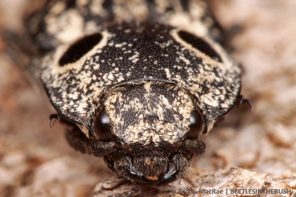
Look into my eye(spot)s!

Look into my eye(spot)s!

If either of those first two lines of defense don’t work, the beetles exhibit “thanatosis” by lying still with legs and antennae appressed to the body to fool the would-be predator into thinking that they are already dead.
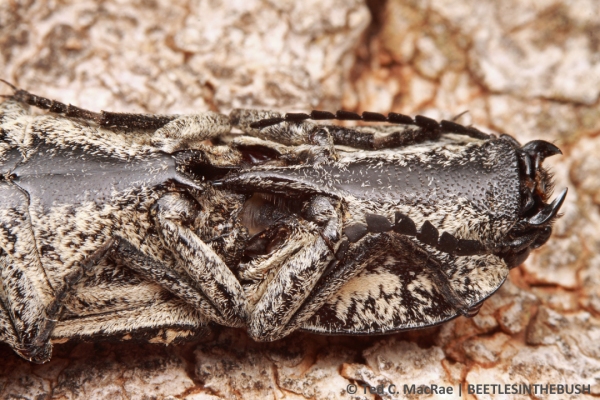
A ventral look at the clicking mechanism between the pro- and mesosterna.

Adults exhibit “thanatosis” (play dead)  when disturbed.

Their most remarkable defensive behavior, however, is their ability to snap or “click” their bodies with enough force to free themselves from the grasp of a novice predator (or careless entomologist). The click is produced by a large prosternal spine and mesosternal notch on the beetle’s underside. To click, the beetle arches back its head and pronotum to retract the spine from the notch cavity, the tip of which is then pressed against the edge of the notch. Muscles within the thorax contract, storing elastic energy, and as the flexible hinge between the pro- and mesothoraces moves, the spine slides until its tip passes over the edge of the notch, releasing the elastic energy stored in the thoracic musculature and snapping the spine back into the notch cavity with enough force to produce an audible click.
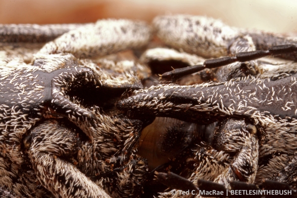
A large spine on the prosternum fits into a groove on the mesosternum.

A large spine on the prosternum fits into a groove on the mesosternum.

This clicking ability also comes in handy if the beetle frees itself from the grasp of a predator and lands on its back. While the beetle’s legs are too short to right itself, its click is capable of launching the beetle high into the air. By tumbling while in the air, the beetle has a 50% chance of landing on its feet (thus, several attempts may be required). When jumping from a hard surface, the beetle is actually capable of launching itself to a height that is several times its body length and can tumble several times while in the air. This raises an interesting question, since theoretically an elevation of only one body length and half of a body revolution are all that is needed for an upside-down beetle to right itself. The power of the click, thus, grossly exceeds the minimal requirement for righting, yet the beetles seem incapable of moderating the force of the click. Furthermore, the 50% probability of landing suggests that they are also incapable of controlling the orientation of their body during the jump and landing. Did the clicking mechanism initially evolve to combat the grasp of predators and was then co-opted for use in jumping, or was the ability to jump the selective pressure that drove its evolution?
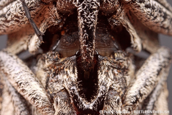
Locked and loaded—the mechanism is primed for the click.

Locked and loaded—the mechanism is primed for the click.

Ribak & Weihs (2011) used biomechanical analyses with Lanelater judaicus to support the idea that the click evolved primarily as a mechanism for vertical jumping. They reason that the excessive vertical distance of the jumps ensures sufficient height when jumping from soft substrates such as foliage or loose soil. A followup study evaluating the effect of natural substrates (Ribak et al. 2012) found that jump height was dramatically reduced (by ~75%) when the beetles jumped from leaves that covered approximately half of the study site and that the reduction in jump height was directly correlated with the amount of work absorbed by the substrate. This provides further evidence that the beetles do not moderate their jumping force and instead simply aim to jump “as high as possible” and rely on random chance for landing back on their feet.
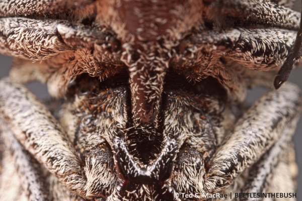
After clicking, the spine returns to its resting position out of the groove.

After clicking, the spine returns to its resting position within the groove.

REFERENCE:

Ribak, G., S. Reingold & D. Weihs. 2012. The effect of natural substrates on jump height in click-beetles. Functional Ecology 26(2):493–499 [abstract].

Ribak, G. & D. Weihs. 2011. Jumping without using legs: The jump of the click-beetles (Elateridae) is morphologically constrained. PLoS ONE 6(6):e20871. doi:10.1371/journal.pone.0020871 [full text].

Copyright © Ted C. MacRae 2014

Out with the old (but on a good note)

This past March was the warmest on record here in Missouri and that made for some nice opportunities to get out and photograph. That said, my enthusiasm for macro photography has been somewhat tempered since my camera body is getting old and showing some signs that it might be on its last leg. The mere fact that I am still using a camera from 2004 may be your first indication that I am something less than a macro photography perfectionist and this is a reflection of the equipment that I first used when I began shooting macro. I started with Canon manual equipment in the mid-1990’s because I thought this would be the best way to learn photography. My stint with a used, and malfunctioning, Canon AE-1 was thankfully short. It was stolen as I returned from a photography trip to the Chiricahua Mountains, but I still lament that the thief made off with the spent rolls of film from the trip! The experience with the AE-1 pushed me towards higher quality, more professional equipment that would stand up to field conditions better. Next up was the Canon F1 and then the wonderfully solid Canon F1N followed later with the game-changing addition of a power winder. The latter was great since insects typically didn’t wait around for me to manually wind the film, refocus, and shoot. The real challenge though was getting the lighting right. At fist I often times had the camera on a tripod which resulted in too many missed shots, restricted what I could shoot, and it didn’t meet what I later realized was my overall goal of macro photography. What I wanted was to have the ability to take satisfactory macro images while not loading myself down with equipment that would detract from also being able to conduct research or simply observe/enjoy nature without trying to capture it on film. I still have a graveyard of old flashes, brackets, and bracket parts that I employed in various combinations to get suitable lighting though it is now clear that I was never able to get the perfect balance. That changed in 2001 when Canon introduced the new MT-24EX macro twin flash and I bought a 1V body. After testing the new system a minimal amount, I realized that macro photography was now made easy relative to what I had wrestled with over the years prior. I had the complete package—no bulky brackets, no hand holding flashes, more certainty of exposure, and quick field set-up. The twin flash allowed me to fire off just the minimal amount of light to obtain my desired depth of field and the flash had sufficient battery power at the lower flash output settings to essentially shoot consecutively without having to wait for the flash to power-up.

Well, at least I thought I had the perfect setup until Ted MacRae took up macro photography and soon demonstrated that options for continuous improvement exist even for this system.

In 2004 I sadly shelved the 1V body and went digital. I briefly used the Canon 10D, which seemed inadequate for various reasons, but then jumped on the newly released Canon 1D Mark II during that same year. It was more than I wanted to spend but there weren’t many options and the benefits were too much to pass up. Most notably, I was sick of buying film, scanning slides, and most of all my hand ached from cataloguing so many slides, i.e., writing a unique code on each slide prior to archiving it in plastic. The time savings alone made the 1D Mark II price palatable and I hoped that investing in a top notch, newly released body would help it remain relevant for a while. I never looked back.

That brings me to that warm day this past March when I settled in to photograph a toad bug, Gelastocoris oculatus (Heteroptera: Gelastocoridae), along the margin of an intermittent creek in Perry Co., Missouri.  As I mentioned above I felt like I was limping along with my dated 1D Mark II but I couldn’t pass up the opportunity to again photograph an individual of G. oculatus that so superbly blended in with its substrate (Figures 1 and 2).

Figure 1. Gelastocoris oculatus, 3.13.2012, Perry Co., MO

Figure 2. Gelastocoris oculatus, 3.13.2012, Perry Co., MO

As I have mentioned before I like to take multiple shots of a subject to help tell a story (yes, including the obligatory head-on close up that Ted always mentions). One important shot in the series can be a photograph from a distance to: 1) better see the subject in its surroundings which can give more insight into its natural history; 2) offer a more artistic view, or, in this case; 3) to show the effectiveness of its cryptic coloration. The actual close-up is great for detail but only gives the viewer an idea that the subject is similarly colored to its background but only a more distant shot really conveys how well the subject melts into the substrate. In this case, Figure 3 was the next progression towards that shot but I was stopped short that day partly due to my middle son falling in the creek and partly due to the apparent malfunctioning of the camera. As you can see, I wasn’t far enough away from the subject to capture what attracted me to the bug in the first place—how well it mimicked its background. In that sense the picture is disappointing because I didn’t finish the story. But on the other hand, the malfunctioning of the camera combined with its age, made it clear that it was now time for a new camera body. So I put on my best frustrated/disappointed face and presented my case to my wife, Jess. It was an easy sell since Jess is… well… at least 95% supportive of my photography. I’ll discuss the new body and the first images soon.

Figure 3. Gelastocoris oculatus, 3.13.2012, Perry Co., MO

Below are more of the variable faces of G. oculatus that I have come across over the years (Figs. 4 – 8). Gelastocoris oculatus is one of two species of Gelastocoris that we have in North America (Arnett 2000). Gelastocoris oculatus can be found continent-wide however G. rotundatus ranges only in the southwest. I love the origin of the family name which the online Merriam-Webster dictionary mentions is from the Greek “gelastos”+ “koris” which translates to “laughable bug”, no doubt due to its odd appearance. Both species are predators that live along the margins of water. Their predaceous nature is made clear by the appearance of their powerful forlegs clearly specialized for catching and securing prey. As you can see, I do not have a picture of their forlegs so I’ve got a great reason to again get down on their level with the new camera.

Figure 4. Gelastocoris oculatus, 5.23.2010, Perry Co., MO

REFERENCE:

Arnett, R.H. 2000. American Insects: A Handbook for the Insects of America North of Mexico. Boca Raton, Florida: CRC Press.

Copyright © Christopher R. Brown 2012