ESA Insect Macrophotography Workshop

Today is the last day of the Entomological Society of America (ESA) Annual Meeting in Austin, Texas, and it has been an action packed week for me. Annual meetings such as this serve several purposes. In addition to seeing talks on a variety of subjects—in my case covering subjects ranging from insect resistance management to scientific outreach to beetle systematics—they also offer the chance to establish new connections with other entomologists that share common interests and reinforce existing ones. Of course, a major part of my interest in entomology revolves around insect macrophotography, and in recent years ESA has begun to cater to the entomological photographer contingent within the society. Last year’s meetings featured a macrophotography symposium titled, “Entomologists Beyond Borders” (for which I was one of the invited speakers), and this year featured an Insect Macrophotography Workshop led by Austin-based entomologist/photographer Ian Wright. Having done this for a few years now I figured a lot of the workshop might be review for me, but I still have much to learn and am willing to accept new ideas from any source. Besides, the workshop involved a field trip to a local habitat to try out our insect photography skills, and for a field junkie like me time in the field at an otherwise all-indoor event spanning close to a week is always welcome. The location of the meetings in Austin this year made this possible, as even in mid-November there still remain insects out and about that can be photographed if the weather cooperates (and it did).

This will be a somewhat different post than what I usually post here. Rather than featuring photos of a certain species and using them as a backdrop for a more detailed look at their taxonomy or natural history, I’m just going to post all the photos that I ended up keeping from the field trip portion of the workshop with just a comment or two about each. We went to the city’s nearby waste-water treatment facility, the grounds of which are wild and woolly enough to provide habitat for insects, and spent about an hour and a half seeing what we could find. For myself, it was a chance to photograph some insects I’ve not normally tried to photograph (i.e., dragonflies, ambush bugs) and get more practice on my blue sky technique. I did appreciate the chance to spend some time talking to Ian during while we traveled to the site and back, and I also ended up helping other participants with their camera equipment questions and technique suggestions. With that, here are the photos I took—I’ll be curious to see what readers think of this post format versus my more typical style.

Micrutalis calva

Micrutalis calva (Hemiptera: Membracidae) on silverleaf nightshade (Solanum elaeagnifolium).

Micrutalis calva

This species of treehopper is restricted to herbaceous plant hosts.

Anax junius

Anax junius (Odonata: Aeshnidae), one of the darner species of dragonfly.

Anax junius

This adult was perched on a dead twig tip and seemed to be “asleep.”

Anax junius

I clipped the perch and held it up for these “in-your-face” shots – it then awoke with a start and flew off.

Phymata sp.

Phymata sp. (Hemiptera: Reduviidae), one of the so-called “jagged ambush bugs.”

Phymata sp.

Formerly a separate family, ambush bugs are now combined with assassin bugs (family Reduviidae).

Acmaeodera flavomarginata

Acmaeodera flavomarginata (Coleoptera: Buprestidae).

Acmaeodera flavomarginata

This is one of a few species of jewel beetle in the southcentral US that are active during the fall.

Mecaphesa sp.

Mecaphesa sp. (Araneae: Thomisidae), one of the crab spiders

Mecaphesa sp.

Cryptic coloration allows the spider to lurk unseen by potential insect prey visiting the flower.

Gratiana pallidula

Gratiana pallidula (Coleoptera: Chrysomelidae) on silverleaf nightshade (Solanum eleagnifolium).

Gratiana pallidula

A type of tortoise beetle, adults “clamp” down against the leaf as a defense against predators.

Copyright © Ted C. MacRae 2013

Bee Assassin on Coneflower

Bee assassin (Apiomerus spissipes) on coneflower (Echinacea sp.) | Gloss Mountain State Park, Major Co., Oklahoma

Apiomerus spissipes? on coneflower (Echinacea sp.) | Gloss Mountain State Park, Major Co., Oklahoma

While looking for longhorned beetles on prickly pear cactus (Opuntia macrorhiza) at Gloss Mountains State Park, I saw a coneflower that didn’t look quite right—there was nothing on the top, but there seemed to be something on the underside. I knelt down cautiously and peered underneath the blossom to find this bee assassin (family Reduviidae, genus Apiomerus) lurking under the petals. As a collector with eyes always looking for signs of insects, I’ve trained myself to look for both the obvious and the non-obvious, yet  this brightly colored insect still almost completely escaped my notice. I can imagine that a bee with little room in its mind for anything but collecting pollen would be easy pickings for such a stealthy predator.

Apiomerus spissipes

The coloration of this individual seems to best match specimens representing the species Apiomerus spissipes, which ranges broadly and abundantly across the Great Plains (Berniker et al. 2011). The location of Gloss Mountain State Park in western Oklahoma places it almost smack in the middle of the recorded distribution for this species, which is largely replaced further east by the closely related but generally darker A. crassipes. Interestingly, very few Oklahoma specimens were available for examination by Berniker and colleagues during their study, a fact that once again demonstrates the need for continued collecting in even “well-collected” states like Oklahoma.

REFERENCE:

Berniker, l., S. Szerlip, D. Forero & C. Weirauch. 2011. Revision of the crassipes and pictipes species groups of Apiomerus Hahn (Hemiptera: Reduviidae: Harpactorinae). Zootaxa 2949:1–113.

Copyright © Ted C. MacRae 2013

Raining spit

Cephisus siccifolia

Cephisus siccifolia 3rd instar nymph | Buenos Aires, Argentina

Even though it was November (and thus spring in Argentina), conditions were already unusually dry—a portent of the worst drought that would hit Argentina in 70 years. Because of this, I found the occasional wet spot on the pavement as I walked the trails in La Reserva Ecológica Costanera Sur rather odd. At first I thought they were spit—the trails were popular on this day for runners and bike riders, but I quickly realized that those would have to be some truly ginormous spit wads based on the size of the splatter. It wasn’t long before I thought to look up, and this is what I saw on the branch directly above me:

Cephisus siccifolia

Cephisus siccifolia spittle mass on unknown species of tree.

I knew right away this was the work of a froghopper, or “spittlebug,” a true bug (order Hemiptera) in the family Cercopidae. Spittlebugs are common in the eastern U.S. where I live and are famous for the spit-like wads of froth (“cuckoo spit” to some) within which the nymphs conceal themselves until they reach adulthood. Our eastern U.S. species, however, are most commonly seen on herbaceous plants rather than in trees, and the frothy masses they produce are fairly small—about the size of a real wad of spit (at least, according to my direct comparison when I was 12 years old). The spittle masses I was seeing today were enormous, frothy, liquid masses that literally dripped from the trees by their own weight—raining spit!

Cephisus siccifolia

Nymphs produce bubbles by siphoning air into a channel under the abdomen.

I was about to move on when I noticed some movement in the spittle mass. A closer look through the macro lens revealed the tip of the abdomen of a nymph slowly circling around near the surface of the spittle and creating new bubbles as it did this. As one can imagine, living inside a mass of bubbly liquid presents a challenge to breathing, and the nymphs get around this problem by protruding the tip of the abdomen outside the spittle mass and taking in air through a tubelike canal below the abdomen (Hamilton & Morales 1992). Strong contractions of the abdomen inside the spittle mass eject air from the canal, resulting in bubble formation.

Cephisus siccifolia

Nymphs partially exposed by removal of spittle mass.

I sent these photos to Andy Hamilton (Canadian National Collection of Insects, Arachnids and Nematodes), specialist in world Cercopidae, to see if there was any chance he might recognize the genus or species based on these photos. I noted that these were the biggest spittlebug nymphs I had ever seen (the individual in the first photo measuring ~10mm in length). Not only did he recognize them as belonging to the genus Cephisus, but he was actually in the process of finishing up a revision of the New World members of the tribe Ptyelini—Cephisus being the sole New World genus to represent the tribe. Based on its white coloration and occurrence as far south as Buenos Aires, Argentina, he suggested this must be C. siccifolia—a species that can sometimes achieve economic pest status (Ribeiro et al. 2005) but which still apparently needs to be properly recorded from Argentina (Hamilton 2012). Based on degree of wing pad development, Andy surmises the individual in Photo #1 represents a 3rd instar (if the 3rd instar measures 10 mm, can you imagine the size of the 5th instars?!). Andy asked me if I would grant him use of the photos in his soon-to-be-published revision (of course I agreed), and here is the plate with the photos (as well as an adult photographed by someone else) as it appears in his paper:

Hamilton_2012_Cephisus

As Andy notes in his paper, it seems rather unusual that Cephisus is the only tribal representative in the New World despite having successfully colonized all of its tropical and subtropical mainland areas. There are several other genera in the tribe in Africa, which would suggest that the Ptyelini arose prior to the late Cretaceous rifting that separated South America and Africa into two continents. It is thus puzzling why the tribe went on to further diversify in Africa but not in the New World.

A tight crop of Photo #3 above was featured in , for which Ben Coulter was the hands-down winner. Honestly I thought this might end up being a slam dunk challenge—people have gotten very good at designing Google search strings to come up with answers that in pre-internet days might have been impossible to find. Nobody stumbled upon the magic search string for this challenge—”MacRae Cercopidae” which pulls up the Hamilton paper and above plate as the very first result. Still, Ben used good old fashioned intuition based on the locality tag to correctly surmise the species and take the early lead in BitB Challenge Session #7. Congratulations, Ben!

REFERENCES:

Hamilton, K. G. A. 2012. Revision of Neotropical aphrophorine spittlebugs, part 1: Ptyelini (Hemiptera, Cercopoidea). Zootaxa 3497:41–59.

Hamilton, K. G. A. & C. F. Morales. 1992. Cercopidae (Insecta: Homoptera). Fauna of New Zealand 25, 40 pages.

Ribeiro, G. T., M. da Costa Mendonça, J. Basílio de Mesquita, J. C. Zanuncio G. S. & Carvalho. 2005. Spittlebug Cephisus siccifolius damaging eucalypt plants in the State of Bahia, Brazil. Pesquisa Agropecuária Brasileira 40(7):unpaginated.

Copyright © Ted C. MacRae 2013

Best of BitB 2012

Welcome to the 5th Annual “Best of BitB”, where I pick my favorite photographs from the past year. 2012 was one of the most intensive travel years I’ve ever had—I spent 8 weeks in Argentina from February through April, made separate trips to Puerto Rico and Arkansas in May (bracketing a personal week in California), traveled almost weekly to Illinois and Tennessee from June to September (interrupted by a personal week in Florida in July), toured the southeastern U.S. (Arkansas, Louisiana, Mississippi and Georgia—great food!) in early September, chased tiger beetles in Oklahoma, Texas and Arkansas in late September, went back to Argentina for a week in October, and capped off the travel year by attending the Entomological Society of America Annual Meetings in Knoxville, Tennessee (for the first time in more than 10 years!)—whew! While many would cringe at such a travel load, I am among the lucky few who actually get paid for doing something that is also my hobby—entomology! This gives me ample opportunity to further hone my photography skills (nine of the 13 photos I’ve selected below were actually taken while I was on business travel), resulting in two key accomplishments this year—my first ever photography talk at the ESA’s insect photography symposium and my first commercial sales (look for the BitB commercial site to go online in 2013).

Enough blather! Here are my favorite BitB photographs from 2012. Click the link in the text below the photo to see the original post. I would greatly appreciate knowing if you have a favorite (and why)—your feedback will be enormously helpful to me as I continue to learn and develop as a photographer.  For those interested, here are my previous year picks for 2008, 2009, 2010 and 2011. And, as always, thank you for your readership!


Spintherophyta (?) sp. in flower of Abutilon pauciflorum | Buenos Aires, Argentina

From  (posted 2 Feb). One of my 2012 learnings was that sometimes a photograph that is not so close is more effective than one that is as close as possible. In one of my earlier attempts at “not-so-close” macrophotgraphy, the soft colors of the flower compliment the brash shininess of the tiny leaf beetle that has been feeding on its pollen. Pink lines lead the eye directly to the subject and create a pleasing composition, and pollen grains stuck to the beetle—a distraction in some situations—add to the miniature natural history story of the photo.


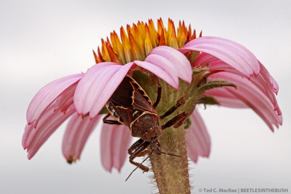
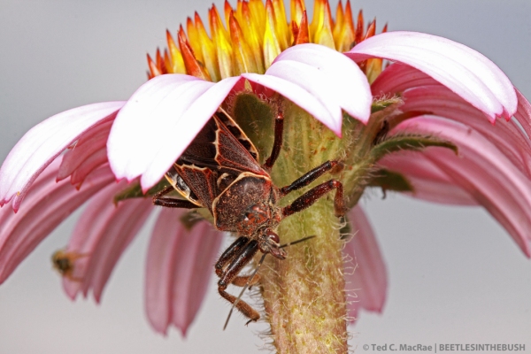
Apiomerus flavipennis with stink bug prey and kleptoparasitic flies | Chaco Province, Argentina

From  (posted 11 Mar). I selected this photo solely for the complex natural history story drama it shows—stink bug (Piezodorus guildenii) feeding on soybean becomes prey of an assassin bug (Apiomerus flavipennis), with volatiles from the chemicals it emitted in a vain attempt to defend itself serving as cues to kleptoparasitic flies (families Milichiidae and Chloropidae) that benefit from the assassin bug’s labors.


Planthopper nymph | Buenos Aires Province, Argentina

From  (posted 26 Mar). Another learning that I began putting into practice in 2012 was the use of low perspective for compositional impact. The cryptic coloration of this planthopper nymph (family Fulgoridae) made it almost invisible on the branch on which it was sitting when viewed from a normal “top-down” human perspective. Getting “down under” it, however, brought the nymph to life and emphasized its unusual form.


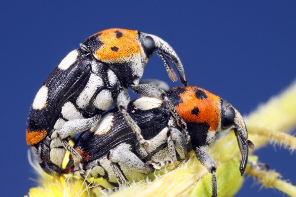
Megabaris quadriguttatus | Corrientes Province, Argentina

From  (posted 12 Apr). I spent much of 2012 working on the “blue sky background” technique, with these weevils from northern Argentina representing one of my better attempts. Macrophotography of insects with a blue sky background involves setting exposure, ISO, and aperture to achieve two separate exposures—full flash illumination of the subject for maximum depth-of-field, and ambient light from the sky to create a clean, uncluttered, natural-looking background. In this shot I managed to achieve an almost ideal shade of blue to compliment the wild black, white and red colors of the beetles. (My one criticism of the photo is having clipped one of the beetle’s feet.)


Bombylius sp. cf. mexicanus | Scott Co., Missouri

From  (posted 16 May). This photo is unusual if nothing else. Focus, lighting, depth-of-field, and composition are all better than can be hoped for in a single shot, but the subject—perfectly alive—is in a most unusual position. Read the original post to find out how this happened.


Perisphaerus sp. (a pill roach) | Vietnam (captive individual)

From  (posted 27 May). White-box photography is an excellent technique for clean, uncluttered photographs of insects, but it also isolates them from their natural surroundings and limits their natural history appeal. The best white-box photos are those that highlight a key feature or behavior of the subject—in this case a pill roach’s comically conglobulating defensive posture.


Micronaspis floridana (Florida intertidal firefly) larva | Pinellas Co., Florida

From  (posted 31 July). Here is another photo whose back story played a big part in its selection. This firefly larva not only represents a rare Florida-endemic species but was also first seen by my then 12-year old nephew, who willingly accompanied me through a dark, spooky salt marsh in the middle of a humid Florida night to see what he could learn. The lesson here for budding natural historians (and old-timers like me) cannot be overstated!


Arctosa littoralis (beach wolf spider) | Lewis Co., Missouri

From  (posted 23 Aug—prelude to  posted 28 Aug). Those who follow this blog know of my obsession with close-up portraits, and while tiger beetles are the subjects I most commonly photograph in this manner, I am always on the lookout for good subjects in other taxa. This wolf spider “face” almost looks human, with “two” eyes, two “nostrils” and a shiny upper lip above huge (albeit hairy) buck teeth! It’s enough fill-the-frame spidery goodness to melt (or explode) the heart of even the most ardent arachnophobe!


Anticarsia gemmatalis (velvetbean caterpillar) egg on soybean leaf

From Life at 8X—Guide to lepidopteran eggs on soybean (posted 3 Sep). “Life at 8X” was a new series I introduced this year, featuring insects photographed at magnifications testing the upper limit of my equipment and photographic skills. Diffraction is the chief difficulty with magnifications as high as this and is the primary flaw in the above photograph. Nevertheless, such view of a moth egg on the underside of a soybean leaf provides a spectacular view of the otherwise unseen micro-world that lives right beneath our noses.


Megacyllene decora (amorpha borer) on snakeroot flowers | Mississippi Co., Missouri

From  (posted 12 Sep). This second example of “blue sky background” was taken later in the year and was considerably more difficult to capture than the first because of the larger size of the subject and resulting need for a longer focal length macro lens. Getting a well-lit, focused, and composed image with a desirable shade of blue in the background depended not only on finding the proper camera settings, but also secure body and camera bracing techniques for this completely hand-held shot.


Cicindelidia politula politula (Limestone Tiger Beetle) | Montague Co., Texas

From  (posted 28 Sep). I will go ahead and say it—this is my favorite photograph of 2012. As discussed under the first entry, panning back from the subject can allow for some very interesting compositions. This photo combines charismatic pose by a wary subject with panning back and low perspective to create an image that scores high in both natural history and aesthetic appeal.


Calosoma sayi (black caterpillar hunter) | New Madrid Co., Missouri

From Black is beautiful! (posted 7 Nov). Of course, close-as-possible can also be used to create striking photos, especially if the subject exhibits features that are best seen up close. Anything with jaws fits the bill in my book, and highlighting the mandibular sculpturing of this caterpillar hunter (a type of ground beetle) required precise angling of the flash heads for maximum effect.


Cicindela repanda (Bronze Tiger Beetle) | St. Louis Co., Missouri

From  (12 Nov). This final selection is not a rare species, but it is as close as I have come to what I consider the “perfect” tiger beetle macrophotograph—a close, low angle, lateral profile of an adult in full-stilt posture (a thermoregulatory behavior), well lit, perfectly focused, and with a dynamic but pleasingly blurred background. It’s a perfect storm of a photo that took the better part of two hours to achieve—rarely do all of these elements come together in a hand-held photograph of an unconfined tiger beetle in its native habitat.


Well, there you have it. I hope you’ve enjoyed my selections, and again please do let me know if you have a personal favorite. See you in 2013!

Copyright © Ted C. MacRae 2012

More Eocene insects

Most of the Green River Formation (GRF) insect fossils that I have on loan clearly represent either beetles (order Coleoptera) or flies (order Diptera). I’ve already shown a few of the latter (fungus gnat, midge), as well as some that don’t belong to either order (ant, cricket?). Here are a few more that seem identifiable to order, but family-level identification is less certain. Thoughts from the readership would be most welcome.


IMG_1923_enh_1080x720IMG_1826_enh_1080x720
This fossil shows an aggregation of insects that I believe represent some kind of beetle. Based on shape and size (16.7 mm length) I’m guessing perhaps either a diving beetle (family Dytiscidae) or whirligig beetle (family Gyrinidae). These are both aquatic families, although only the former is among the beetle families recorded from the GRF by Wilson (1978).


IMG_1927_enh_1080x720 IMG_1836_enh_1080x720
There are two insect fossils on this specimen, but the closeup is the one near the center of the rock. It is tiny (3.5 mm in length), and at first I thought it might be a fly (order Diptera). However, dipterist Chris Borkent thinks it might be a small hymenopteran (bee?) because it has what looks to be long multi-segmented antennae. The only bee family recorded for the GRF by Wilson (1978) is Anthophoridae (now included within Apidae), of which this fossil clearly is not a representative. There are six other hymenopteran families recorded in that work, of which Tenthredinidae is the only one that seems plausible. Of course, it could represent a family not recorded by Wilson (1978). Collected along Hwy 139 in Douglas Pass (Garfield Co., Colorado).

Here is a closeup of the other fossil (far right in photo above). This looks to me like a brachyceran fly, and I’ve sent a high resolution version of the image to Chris Borkent to see what he thinks.


IMG_1934_enh_1080x720 IMG_1850_enh_1080x720
The label accompanying this fossil indicates “Mosquito (?),” but to my eye this looks like a true bug (order Hemiptera). It is small—only 5.9 mm in length—and has the gestalt of a plant but (family Miridae) or seed bug (family Lygaeidae). GRF fossils representing the latter but not the former were recorded by Wilson (1978). Also collected along Hwy 139 in Douglas Pass (Garfield Co., Colorado).


REFERENCES:

Wilson, M. V. H. 1978. Paleogene insect faunas of western North America. Quaestiones Entomologicae 14(1):13–34.

Copyright © Ted C. MacRae 2012

Life at 8X: MPMI Cover


The January 2013 issue of Molecular Plant-Microbe Interactions (volume 26, number 1) is now online. Why do I mention this? You may recall the cover photos of the soybean aphid, Aphis glycines, from my post —one of a series of posts I’ve done featuring insects photographed at 8X life-size.

MPMI is a publication of The American Phytopathological Society, and I have Dr. Gustavo MacIntosh at Iowa State University to thank for the appearance of these photos on the cover of this Special Focus Issue. Dr. MacIntosh is Associate Professor of Biochemistry, Biophysics and Molecular Biology and studies hormone-based defense mechanisms in soybeans. In a paper appearing in this special issue, Dr. MacIntosh and co-author Matthew Studham published the results of a study that suggests soybean aphids are able to “short-circuit” soybean defense mechanisms, making it easier for other pests (e.g., soybean cyst nematode) to colonize infested plants as well. Their study revealed large differences in transcription profiles of soybean varieties with and without an endogenous resistance gene (Rag1) in response to aphid infestation and suggested that the aphids are able to circumvent the defense response in susceptible plants by triggering activation of abscissic acid (normally associated with abiotic stress responses) as a “decoy” strategy (Studham & MacIntosh 2013). Plants infested with aphids have been shown to also become more susceptible to soybean cyst nematode—even varieties with genetic resistance to nematodes (McCarville et al. 2012). Dr. MacIntosh saw my photos when I posted them here and asked permission to submit them as candidates for the cover of the MPMI issue in which his paper was to appear.

Dr. Macintosh hopes that his research will enable the development of soybean varieties that will be more resistant to aphids and other pests.

REFERENCE:

McCarville, M. T., M. O’Neal, G. L. Tylka, C. Kanobe & G. C. MacIntosh. 2012. A nematode, fungus, and aphid interact via a shared host plant: implications for soybean management. Entomologia Experimentalis et Applicata 143(1):55–66 [DOI: 10.1111/j.1570-7458.2012.01227.x].

Studham, M. E. & G. C. MacIntosh. 2013. Multiple Phytohormone Signals Control the Transcriptional Response to Soybean Aphid Infestation in Susceptible and Resistant Soybean Plants. Molecular Plant-Microbe Interactions 26(1):116–129 [DOI: 10.1094/MPMI-05-12-0124-FI].

Copyright © Ted C. MacRae 2012

Life at 8X—Bandedwinged Whitefly

Trialeurodes abutiloneus (bandedwinged whitefly) | Obion Co., Tennessee

The world of minute insects can seem strange and even bizarre when compared to our relatively giant perspective. To the unaided human eye, this bandedwinged whitefly (Trialeurodes abutiloneus), measuring only 1 mm in length, looks like nothing more than a fleck of dandruff. Through a Canon MP-E 65mm 1–5X macro lens with 68 mm of extension tube (resulting in 8X magnification), however, we see an almost moth-like insect with a decidedly adorable “face” negotiating the “trichome forest” of a soybean leaf under-side.

A more conventional 2X view of a whitefly infestation on the underside of a leaf

Whiteflies (order Hemiptera, family Aleyrodidae) are tiny insects (more related to aphids than true flies) that colonize a variety of host plants, often building to extraordinary numbers and densities while sucking juices from the leaves. The bandwinged whiteflies in these photos were seen in a soybean field in northwestern Tennessee this summer and can be easily identified as this species due to the transverse, zig-zag bands on the forewings (Malumphy et al. 2010). In the photo above numerous eggs can also be seen distributed over the leaf surface—a sign that this population is about to explode given the numbers of eggs present.

Zooming in to 8X allows the zig-zag wing pattern to be seen easily.

Whiteflies are an occasional pest of soybean in the U.S., but yield reduction has been documented only in the southeastern U.S. by another species, Bemisia tabaci (sweet potato whitefly). Whiteflies are also occasionally seen on soybeans in the Midwest by B. tabaci or yet another species, Trialeurodes vaporarium (greenhouse whitefly); however, yield impacts in this area are rare. Trialeurodes abutiloneus is occasionally reported from soybean, but this species is actually more commonly encountered on sweet potato and malvaceous crops such as cotton and hibiscus (Clower et al. 1973). There was a lot of cotton growing in the area of this soybean field, so perhaps this infestation was a result of spillover from that crop.

Piercing/sucking mouthparts are inserted into the leaf for feeding.

“Adorable” and “cute” are not words that I’ve ever associated with whiteflies, but these ultra-closeup photographs give them a personality that I’ve not seen before. For an even more astounding view of the face of a greenhouse whitefly, see this incredible 16X photograph by Huub de Waard. Taken with the same lens as these—though I suspect with a 2X converter rather than extension tubes, it shows an amazing level of sharpness compared to the admittedly soft photos in this post. The larger aperture used (f/6.3) may also be a better choice than the small f/13 aperture I used in an attempt to preserve as much depth of field as possible but with which diffraction is likely significantly greater. Stay tuned as I do some more testing…

A cute couple!

REFERENCE:

Clower, D. F. & C. M. Watve. 1973. The bandedwinged whitefly as a pest of cotton, pp. 90–91. Proceedings of Beltwide Cotton Production and Research Conference, 11–12 January, Phoenix, Arizona. Cotton Council of America, Memphis, TN.

Malumphy, C., A. MacLeod & D. Eyre. 2010. Banded-winged whitefly Trialeurodes abutiloneus. Plant Pest Factsheet, The Food and Environment Research Agency (Fera), 4 pp.

Copyright © Ted C. MacRae 2012

Life at 8X—soybean aphid

Although my first attempt at adding extension tubes to my Canon MP-E 65mm macro lens, effectively converting it from a 1–5X to a 1.7–8.0X lens, was nearly a year ago, it has only been recently that I’ve actually started experimenting with this combination to obtain high-mag photographs of very small insects in the field. The first example that I showed of such a photograph was a tiny seed weevil (Althaeus sp.) on its hibiscus host plant. I’ve since photographed a number of other insect subjects at high-mag using this setup and am getting a better feel for the capabilities—and limitations—inherent in using it. First, here is what the setup actually looks like:

Canon 50D body, MP-E 65mm macro lens on 68mm extension, MT-24EX twin flash w/ DIY diffuser.

Not the normal photo quality for this site (just a quick field setup photographed with my I-Phone), but it shows just how long the lens component becomes when fully extended to achieve 8X magnification. The camera is quite front-heavy, making the camera difficult to use hand-held, and the very shallow DOF (depth of field) due to the extreme level of magnification makes precise focusing difficult and magnifies the effect of any motion between the camera and subject. Obviously, one solution for these problems is to mount the camera on a tripod and place the subject on a stable surface; however, for reasons I’ve mentioned elsewhere, it is unlikely that I will ever take to bringing a tripod into the field, and the whole point of this exercise is to develop the capability for getting usable hand-held field photographs no matter what level of magnification they may require. As an alternative, I use a number of other techniques, discussed in my prior post on the subject, to stabilize the camera without using a tripod.

One of the recent subjects I photographed with this setup is the soybean aphid, Aphis glycines (order Hemiptera, family Aphididae). This distinctive Asian species has recently established in the U.S. as invasive pest of soybeans; adult females measure only 1–2 mm in length (and the nymphs are even smaller) and can quickly develop very high densities on the leaves and upper stems of soybean plants. The following photograph was taken at the camera setup’s minimum magnification of 1.7X and provides a typical view of adult aphids and their progeny:

Aphis glycines (soybean aphid) | Warren Co., Illinois

While the above photograph does a very good job of showing the colonial appearance of infestations by these aphids on soybean foliage, what about the aphids themselves? It would be nice to get a better look at individual aphids. The following photographs were all taken with the lens fully extended to achieve 8X magnification (and completely hand-held):

Adult female aphid—note the eye spots of the unborn nymphs visible within the body.

Another adult female navigates the hairs on the surface of the soybean leaf (I never knew soybean leaves were so hairy!).

A mother surrounded by her progeny. As above, eye spots of unborn nymphs can be seen inside her body.

These photographs are not without their problems—they are a bit soft, probably due to motion blur that results from the camera being hand-held and the extremely thin DOF that makes it difficult to get all of the desired components of the photos equally in focus. Lighting also is a challenge, as the very small subject-to-lens distance forces light from the flash to come from directly above or even behind the subject while minimizing front lighting (especially evident in the last photo with its straight down view). Nevertheless, these are decent, usable photographs that provide an uncommon view of these exceedingly tiny insects—without the encumbrance of carrying a tripod in the field, the time investment of studio photography and/or focus-stacking, or the expense of a microscope-mounted camera system (for those of us without access to such systems).

Copyright © Ted C. MacRae 2012