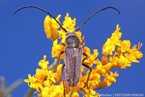
Mallodon dasystomus | southeast Missouri (Mississippi Co.)
Today’s (slightly belated) edition of “One-shot Wednesday” features a beetle that I saw just about this time last year while blacklighting along the Mississippi River in the southeastern lowlands of Missouri. Mallodon dasystomus¹ is a prionid longhorned beetle (family Cerambycidae, subfamily Prioninae) that is sometimes called the “hardwood stump borer”. It is perhaps one of the most widely distributed members of its group, occurring across the southern tier of the U.S. down through Mexico and Central America as far as northern South America.
¹ Until recently the specific epithet was consistently misspelled in most of the literature as “dasytomus“. A closer look at the Greek root words dasus (δασύς), meaning “hairy”, and stoma (στόμα), meaning “mouth”, shows the misspelling to be nonsensical. I, myself, am guilty of using the wrong spelling in my checklist of Missouri longhorned beetles (MacRae 1994), although I can claim to have been “going with the flow”.
Despite the beetle’s wide geographical range, I searched for it both eagerly and unsuccessfully during the 1980s as I was gathering data for my checklist of Missouri longhorned beetles (MacRae 1994). I eventually published that checklist and included the species on the basis of a few specimens seen in other collections, but I never encountered it for myself until some years later during a visit to Cave Creek Canyon in southeast Arizona. As noted by Linsley et al. (1961), this species is common there and is associated with large, partially dead Arizona sycamores (Platanus wrightii). Although nearly 20 years ago, I still recall seeing the large beetles crawling high up on the trunks and sitting in their emergence holes with only their massively-mandibled heads protruding as they tantalizingly waved their antennae about.
That experience would directly lead to my eventually finding this species for myself in Missouri. Shortly after returning to the state in the mid-90s, I was driving along a road in the state’s southeastern lowlands when I passed a very large, half-dead American sycamore (Platanus occidentalis). Even at a speed of 40 mph I could see the large emergence holes that immediately reminded me of what I had seen in Arizona, so I hit the brakes, made a quick U-turn, and came back to look at the tree a little more closely. I was convinced the holes were made by this species, and my hunch was proven when I eventually found several beetle carcasses on the ground around the base of the tree. I returned the following weekend with a chainsaw, cut several one-cubic-foot sections of wood from the dead portion of the massive tree’s trunk (with landowner permission), and eventually reared a nice series of adults from the wood. Having uncovered the association of this species with sycamore in the state, I was able to find the species also in several other locations in southeastern Missouri, but I have not managed to find the species in any areas north of the southeastern lowlands in Missouri despite the common occurrence of the host tree.
The beetle in the above photograph landed on the foliage of a large silver maple (Acer saccharinum) next to the ultraviolet light I had setup in wet bottomland forest along the Mississippi River, and wanting to ensure that I got at least one in situ photo of the beetle that is where I shot it. I did try to move it to the trunk of a large, dead sycamore nearby for a more realistically representative photo of how these beetles are usually encountered, but the beetle became quite agitated when I moved it and my considerable patience was never rewarded. I popped it into a vial in hopes of photographs the next morning, but conditions were not to the beetle’s liking and it expired before I had another chance to photograph it. Just the other night I setup a blacklight in a spot not too far from where I saw this beetle in hopes of getting another chance to photograph it. That effort was not successful, but I did find a longhorned beetle species that I had not seen in nearly 30 years! I was successful in photographing that species but (please excuse the teaser) will save those photos for a future post.
REFERENCES:
Linsley, E. G., J. N. Knull & M. Statham. 1961. A List of Cerambycidae from the Chiricahua Mountain Area, Cochise County, Arizona (Coleoptera). American Museum Novitates 2050:1–34 [full text, pdf].
MacRae, T. C. 1994. Annotated checklist of the longhorned beetles (Coleoptera: Cerambycidae and Disteniidae) known to occur in Missouri. Insecta Mundi 7(4) (1993):223–252 [pdf].
© Ted C. MacRae 2014